


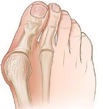
The valgus deformation of the thumb of the foot is the most frequent deformation of the foot, characterized by the deviation of the thumb out.
The disease is bilateral in nature, mainly affecting women over 35 years old.In most cases, the deformation is accompanied by the chronic inflammatory condition of the joints and the joint deformation of the Plusnephalang joints, the pronunciation deviation of the first metatarsal bone.
Causes of the disease.Why is it dangerous?
The causes of the Valgus deviation of the thumb of the feet have not been fully studied.There are some theories about the appearance:
- Theory of relics.In the middle of the 19th century, it was believed that only models were subjected to this deformation due to the use of model shoes in high modes, but in the study, this pathology was found in flat shoes.
- The theory of main muscle weakness - was rejected in a detailed study.
- The theory of the weakness of the ligament movement and the Aponeurosis of the only - most scientists comply with this theory.
There are several factors leading to the launch of this pathology:
- Excess weight increases the load on the foot.
- Change of age -related disorders in the ligament joint apparatus.
- The high heels are more than 5 cm, narrowed into toes.
- The current deformation of the skeleton (scoliosis, Valgus deformation of the femur and knee joint, flat feet).
The danger of the pathology lies in this deformation time leading to the deformation of the posture, which can develop the depth of the spine, hips and knee joints due to inappropriate distribution during walking and muscle injury.
Clinical manifestations
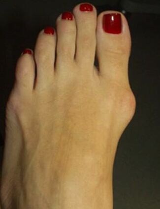
At the beginning of the disease, Valgus deformation is not clinically shown.Most women are more interested in the appearance of the feet-an increase in the Metacarpal-Phalanx joint and the explosion of the first finger becomes noticeable.
Over time, the pain occurs, first when wearing narrow shoes and walking for a long time, and then the pain starts to constant, pain.
Due to the inappropriate weight distribution, the first and second fingers in the base, formed by corn, second fingers were raised, expanded a lot, the fingers of Malleus Malleus were formed, complicated a lot when walking.Due to the decline in blood circulation and conservation on the front of the foot, arthritis and chronic arthritis developed.
Diagnose
- In the process of objective inspection, classic deformation with the Valgus deviation of the thumb can be visible, the far part of the foot has been expanded.In the projection of the first methatarsal bone tip, there are signs of burgous - hypertension and edema of the skin, pain during touch.
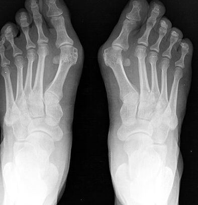
- X -ray of the foot in two projections.The front projection determines the degree of the thumb, the state of the metacarpal-phalanx joint, the level of sesame bone movement and in the side projection, the level of the flat foot is visualized and calculated, often occurs with Valgus deviation of the first finger.
- Plotography.On the foot of the foot, made on paper, a line is drawn through the center of the heel and between the fourth and third fingers, the outer set of the foot is formed in the sense of the ball, whereby the presence of flat feet and its degree is evaluated.In these patients, a flat foot or flat foot of level 1 is more often detected.
Treatment and prevention
Depending on the severity of the process, conservative treatment or surgery is performed.
Conservation treatment is performed in the mild stage of the disease, many different types of orthopedic bases are used, selected individually and bringing deformed fingers to normal positions, while the load in the front of the expenditure is stable and distributed.
In children and old age, the crossroads of the limb with the lying between the first and the second finger are used.To relieve pain, warm bath with sea salt and soda used, radon bathtub for good results.
To prevent Burs inflammation, local anti -inflammatory gel, compressed with dimexide and capocaine used, and with distinct pain, Novocaine blockade and intracellular glucocorticoids are used.
Surgical treatment is possible at any stage of the disease, it is performed under local anesthesia, helping to reduce the list of surgical contraindications.
In phase 1, the cold growth of the bone is removed on the inner edge of the bone, while it can only reduce the pain of the process, but does not prevent the development of deformation in the future.
With the serious deviation of the finger with the presence of flat feet, the removal of the bone is done at the bone of the first finger to use the bone tongue to reject the finger to the normal position, with the help of the lavsan tape, the horizontal ligament of the base is formed.After the surgery, the legs are closely fixed with the tape for 4 weeks, so the individual orthopedic base should be used in the year.
Prevention is to use comfortable shoes without high heels, shoes should sit on the feet freely, a narrow toe should not bring discomfort in the thumb.If there is any deformation of the skeleton, undergoing prevention tests at the orthopedic doctor to identify and slow down the progression of Valgus deformation of the thumb of the feet.
Consequences and complications
A complication of the process is the development of Deichländer or March of the feet, characterized by acute pain, due to the microcracks in the joints of the foot and tendonitis.
Due to continuous inflammation and mechanical damage, there is a risk of developing bone malignant tumors.